Anatomy of Heart And Valves
About the heart
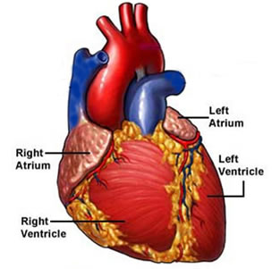
The heart is the hardest working muscle in the human body. Located almost in the center of the chest, the adult human heart is about the size of two fists held side-by-side.
At an average rate of 80 times a minute, the heart beats about 115,000 times in one day or 42 million times in a year. During an average lifetime, the human heart will beat more than 3 billion times - pumping an amount of blood that equals about 1 million barrels.
How the heart works
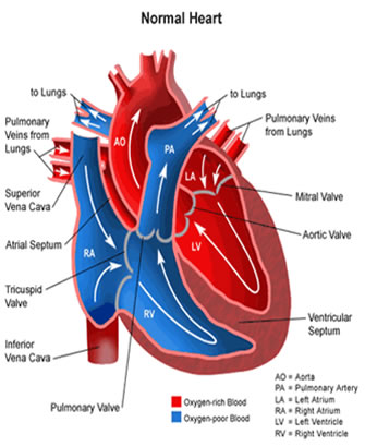
The cardiovascular system, composed of the heart and blood vessels, is responsible for circulating blood throughout your body to supply the body with oxygen and nutrients.
It is made up of:
Four chambers (two atria and two ventricles) that receive blood from the body and pump out blood to it.
The atria receive blood coming back to the heart.
The ventricles pump the blood out of the heart.
Blood vessels, which compose a network of arteries and veins that carry blood throughout the body.
Arteries transport blood from the heart to the body tissues.
Veins carry blood back to the heart.
Four valves to prevent backward flow of blood.
Each valve has flaps, called leaflets,that allow the forward flow of blood and prevent the backward flow.
An electrical system of the heart that controls how fast it beats.
The Conduction System
Electrical impulses from heart muscle (the myocardium) cause heart to contract. This electrical signal begins in the sinoatrial (SA) node, located at the top of the right atrium. The SA node is called the heart's "natural pacemaker." An electrical impulse from this natural pacemaker travels through the muscle fibers of the atria and ventricles, causing them to contract. Although the SA node sends electrical impulses at a certain rate, your heart rate may still change depending on physical demands, stress, or hormonal factors.
The Circulatory System
Your heart and circulatory system make up your cardiovascular system. Your heart works as a pump that pushes blood to the organs, tissues, and cells of your body. Blood delivers oxygen and nutrients to every cell and removes the carbon dioxide and waste products made by those cells. Blood is carried from heart to the rest of the body through a complex network of arteries, arterioles, and capillaries. Blood is returned to heart through venules and veins. If all the vessels of this network in body were laid end-to-end, they would extend for about 60,000 miles (more than 96,500 kilometers), which is far enough to circle the earth more than twice!
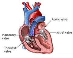
What are heart valves?
The heart consists of four chambers, two atria (upper chambers) and two ventricles (lower chambers). There is a valve through which blood passes before leaving each chamber of the heart. The valves prevent the backward flow of blood.
These valves are actual flaps that are located on each end of the two ventricles (lower chambers of the heart). They act as one-way inlets of blood on one side of a ventricle and one-way outlets of blood on the other side of a ventricle. Each valve actually has three flaps, except the mitral valve, which has two flaps.
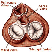
The four heart valves include the following:
- Tricuspid valve: located between the right atrium and the right ventricle.
- Pulmonary valve: located between the right ventricle and the pulmonary artery.
- Mitral valve: located between the left atrium and the left ventricle.
- Aortic valve: located between the left ventricle and the aorta.
How do the heart valves function?
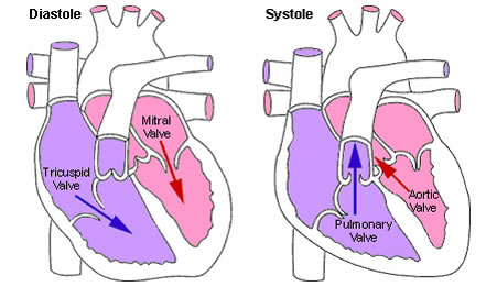
As the heart muscle contracts and relaxes, the valves open and shut, letting blood flow into the ventricles and atria at alternate times.
The following is a step-by-step illustration of how the valves function normally in the left ventricle:
- After the left ventricle contracts, the aortic valve closes and the mitral valve opens, to allow blood to flow from the left atrium into the left ventricle.
- As the left atrium contracts, more blood flows into the left ventricle.
- When the left ventricle contracts again, the mitral valve closes and the aortic valve opens, so blood flows into the aorta.
What is heart valve disease?
Heart valves can have one of two malfunctions:
Regurgitation
The valve(s) does not close completely, causing the blood to flow backward instead of forward through the valve.
Stenosis
The valve(s) opening becomes narrowed or does not form properly, inhibiting the flow of blood out of the ventricle or atria. The heart is forced to pump blood with increased force in order to move blood through the stiff (stenotic) valve(s).
Heart valves can have both malfunctions at the same time (regurgitation and stenosis). When heart valves fail to open and close properly, the implications for the heart can be serious, possibly hampering the heart's ability to pump blood adequately through the body. Heart valve problems are one cause of heart failure.
Causes
- A weakening of the valve tissue caused by energy changes in the body. This is called myxomatous degeneration. It happens most often in elderly patients and commonly affects the mitral valve.
- A buildup of calcium on the aortic or mitral valves, which causes the valves to thicken. This is called calcific degeneration.
- An irregularly shaped aortic valve or a narrowed mitral valve. This is usually a congenital defect, which means that most people who have it were born with it.
- Use of the anti-obesity medicines fen-phen and Redux, which were removed from the market after being linked to heart valve disease.
- An infection in the lining of the heart's walls and valves (the endocardium). This is called infective endocarditis.
- Coronary artery disease.
- Heart attack.
Symptoms
Symptoms depend on the patient and the type and severity of valve disease. Some patients have no symptoms at all. In other cases, valve disease may take its toll over many years. In time, patients may develop congestive heart failure. Also, valve disease may lead to heart muscle disease (cardiomyopathy), an irregular heartbeat (arrhythmia), and blood clots.
Diagnosis
Your doctor can tell if you have valve disease by listening to your heart with a stethoscope. Doctors can listen for the distinct clicking sounds or murmurs of valve disease. Other tests your doctor may order include
- A chest x-ray, which can show if your heart is enlarged.
- Echocardiography, which can show the thickness of your heart's walls, your valves' shape and action, and the size of your valve openings. Doppler echocardiography (ultrasound) can be used to find out how severe the narrowing (stenosis) or backflow (regurgitation) of blood is.
- Electrocardiography (EKG or ECG) can be used to find out if your ventricles or atria are enlarged. This test can also determine if you have an irregular heartbeat (arrhythmia).
- Coronary angiography It lets doctors see your heart as it is pumping. Angiography can help identify a narrowed valve or any backflow of blood.